
Modelling Particle Fracture in 3D using Finite Element (FE) Method
Modelling fracture behavior of individual particles
3D SMT images of particles were used to generate the FE mesh to peform FE modelling of particle fracture

Modelling force transmission and fracture behavior of sand particles within specimens
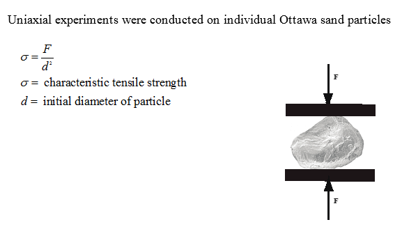
Cil et al. (2017) conducted 1D compression experiment on a uniform natural silica sand known as F-75 Ottawa sand where only grain sizes between US sieve #40 (0.420 mm) and sieve #50 (0.297 mm) were used in the experiment. Sand particles were deposited inside a thick-walled acrylic mold with an inner diameter of 1 mm. The specimen had an initial height of about 2 mm. The experiment was conducted at beamline 1-ID of the Advance Photon Source (APS), Argonne National Laboratory (ANL), Illinois, USA. A special apparatus was used in which both 3D X-ray diffraction (3DXRD) and 3D SMT scans were acquired at different loading stages. The combination of 3DXRD and SMT techniques offers a unique approach to track individual particles and measure their lattice strains simultaneously.
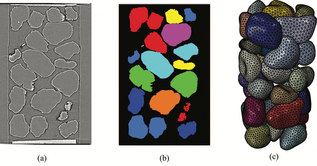
The initial SMT image was used in this paper to develop the FE model that captures the exact shape of sand particles. The initial SMT image was processed using AVIZO Fire 9.3 software in which 35 particles were identified as separate objects and numerical labels (1-35) were assigned to each particle. AVIZO Fire 9.3 software was then used to crop each particle individually and its surface mesh was then generated and saved as stereo-lithography (STL) file producing 35 STL files (one for each particle). A few smoothening and simplification cycles were applied on each surface image to generate a surface mesh with approximately 800 triangles per particle.

Discrete Element Modelling of Sand Behavior
Particle Fracture Behaviour
- PFC3D Software was used in the analysis
- Each particle was modeled as an agglomerate of a large number of non-uniform small sub-spheres bonded together according to the Bonded Particle Model (BPM) of Potyondy & Cundall (2004)
- The sub-spheres are bonded at their contact points by parallel bonds that act as a cementing material.
- The bond strength was chosen randomly from a normal distribution function defined by a mean (475 MPa) and standard deviation (175 MPa) values.

mm agglomerates of sub-spheres with
(b) Rmin = 0.035 mm; (c) Rmin = 0.045 mm; (d) Rmin = 0.035 mm and
non-spherical shape.

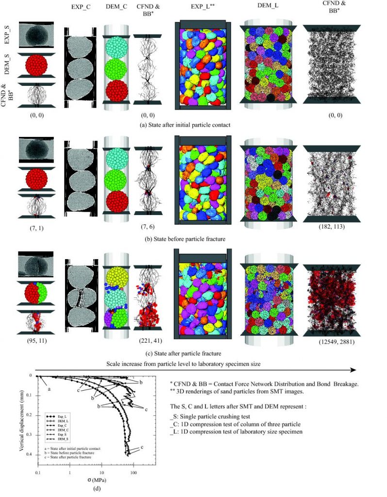
as (tensile, shear). (d) shows a comparison of responses of experiments and DEM simulations for single particle, column of particles and laboratory size specimen.
Related Publications:
- Amirrahmat, S., Imseeh, W., Alshibli, K. A., Kenesei, P., Jarrar, Z., and Sharma, H. (2020). “3D Experimental Measurements of Evolution of Force Chains in Silica Sand”, ASCE Journal of Geotechnical & Geoenvironmental Engineering, Vol. 146, No. 5 (May), https://doi.org/10.1061/(ASCE)GT.1943-5606.0002241
- Amirrahmat, S., Alshibli, K. A., Melton, J. L. (2019). “3D Finite Element Modelling of Fracture of Sand Particles Subjected to High Strain Loading Rate”, Journal of Dynamic Behavior of materials, Vol. 5, Issue 4, pp. 444-462, https://doi.org/10.1007/s40870-019-00211-0
- Druckrey, A., Alshibli, K. A., Casem, D., and Huskins, E. (2018). “Experimental Fracture Analysis of Individual Sand Particles at High Loading Rates”, ASTM Geotechnical Testing Journal, Vol. 41, No. 3, https://doi.org/10.1520/GTJ20170087
- Imseeh, W. H. and Alshibli, K. A. (2018). “3D Finite Element Modelling of Force Transmission and Particle Fracture of Sand”, Computers and Geotechnics, Vol. 94, pp. 184-195, published online September 22, 2017,DOI: https://doi.org/10.1016/j.compgeo.2017.09.008, Download the paper
- Cil, M. B. and Alshibli, K. A., Kenesei, P. (2017). “3D Experimental Measurement of Lattice Strain and Fracture Behavior of Sand Particles Using Synchrotron X-ray Diffraction and Tomography”, ASCE Journal of Geotechnical & Geoenvironmental Engineering, Vol. 143, No. 9,DOI: http://ascelibrary.org/doi/10.1061/%28ASCE%29GT.1943-5606.0001737, Download the paper
- Druckrey, A. M. and Alshibli, K. (2016). “3D Finite Element Modeling of Sand Particle Fracture based on in situ X-Ray Synchrotron Imaging”, International Journal for Numerical and Analytical Methods in Geomechanics, Published online June 4, 2015, Vol. 40, pp. 105-116, DOI: 10.1002/nag.2396
- Cil, M. B. and Alshibli, K. (2014). “3D Evolution of Sand Fracture Under 1D Compression”, Geotechnique, Vol. 64, No. 5, pp. 351-364, http://www.icevirtuallibrary.com/doi/10.1680/geot.13.P.119
- Cil, M. B. and Alshibli, K. A., McDowell, G. R., and Li, H (2013) “Discussion: 3D Assessment of Fracture of Sand Particles using Discrete Element Method”, Geotechnique Letters, Vol. 3, pp. 13-15, DOI: http://dx.doi.org/10.1680/geolett.13.00004
- Cil, M. B. and Alshibli, K. A. (2012) “3D Assessment of Fracture of Sand Particles using Discrete Element Method”, Geotechnique Letters, Vol. 2, pp. 161-166, DOI: http://dx.doi.org/10.1680/geolett.12.00024